

Past projects

C. elegans worm intestines
2026
Red symbiont bacteria inside of green outlined intestines
2025
Squash bug crypts
Early colonization of squash bug gut, green bacterial symbionts, magenta bug actin.
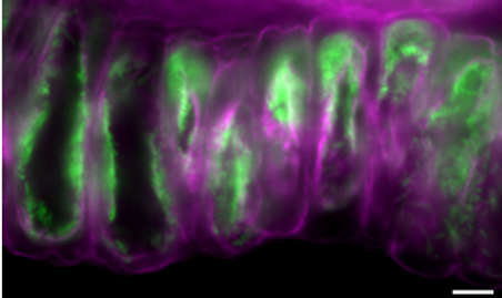

2022
Squash bug crypts (M4)
Here is the larval (L2) intestine of a squash bug with Actin (green) forming crypts and Caballeronia bacteria (magenta) colonizing those newly made crypts.
2021
Squash bug intestine (M4)
Here is the larval (L3) intestine of a squash bug with DNA (Blue), Actin (green), and Caballeronia bacteria (red).

_tif.png)
2021
Filamentous fungi
Here are some fungal hyphae (Basidiomycota) stained for chitin (red) at 20X magnification.
2021
Hemipteran intestinal crypts
Collaborative project with Jason Chen (Gerado/Vega labs, Emory University). This is a 60X magnified view of Symbionts (Caballeronia- Red) inside a squash bug's crypts (actin-green and DNA-White).
_tif.png)

2021
Fungi feeling each other out
Here is my first pictures of parasitic fungus (genus Escovopsis: bottom left) interacting with fungi isolated from the fungal farms of ants (Cyphomermex Mulleri).
2021
Resident farming ants
Here is a short video of Trachymermex ants tending to their fungus garden. They have been living in the lab for a few months. There is supposed to be a queen hiding somewhere in here!

2019
The oral appratus
This is a fluorescence image of Tetrahymena thermophila microtubules and basal bodies converted into a heatmap.

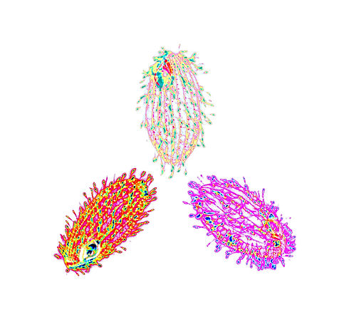
These are the different strains of Tetrahymena thermophila stained for basal bodies (centrioles) and microtubules. Each cell's fluorescence signal was then converted into a different type of heatmap.
I made this art to signify diverse perspectives, orientations, and life histories.
Fun fact: T. thermophila has 7 sexes (mating types).